

X-Rays are an important tool for diagnosing different clinical and dental issues because of their exceptional capacity to give definite pictures of internal structures, including bones and teeth. In dentistry, X-beams assume a significant part in diagnosing dental issues since they offer a few benefits. To begin with, X-rays can uncover issues that are not apparent to the unaided eye, like depressions between teeth, dental abscesses, and impacted wisdom teeth. They additionally empower dental specialists to survey the soundness of the jawbone and recognize conditions like periodontal sickness or bone loss. However, one should Learn the Types and Misconception About Dental X-Rays. Because they are key devices for dental experts, supporting precise determination and compelling treatment and anticipating a large number of dental issues.
What Is It?
Dental Panoramic X-Rays in Dubai, otherwise called dental radiographs, are indicative pictures of a patient’s teeth and encompass oral designs. These pictures are caught utilizing X-beam innovation, which utilizes a modest quantity of ionizing radiation to make comprehensive photos of the teeth, jawbone, and other oral tissues. These are fundamental devices in dentistry since they give important data that is not noticeable during a normal dental assessment.
They are a fundamental piece of routine dental considerations, as they permit dental specialists to recognize and analyze different oral circumstances, including cavities, gum diseases, contaminations, growths, and primary issues. They likewise assume an imperative part in treatment arranging and observing the improvement of dental circumstances. To guarantee patient well-being, dental X-beams are commonly low in radiation dose, and medical professionals keep rules to limit radiation openness while getting the fundamental diagnostic data.
Types:
Dental X-rays encompass several types of radiographic images. These X-rays detect different places in the oral cavity due to which they are differently categorized. It helps to assess and diagnose several oral issues. Dentists choose a certain type according to the condition of the patient.
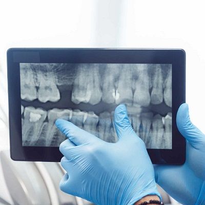
Bitewing X-Rays:
These center around the crowns of the upper and lower back teeth, giving a perspective on the areas between teeth. Bitewing X-beams usually help to identify cavities between teeth, assess the fit of dental fillings, and screen the gum infection.
Periapical X-Rays:
They catch a definite picture of a couple of explicit teeth, including the whole tooth, from crown to root tip. Dental specialists utilize these X-beams to analyze issues like tooth breaks, contaminations, root anomalies, and changes in tooth structure.
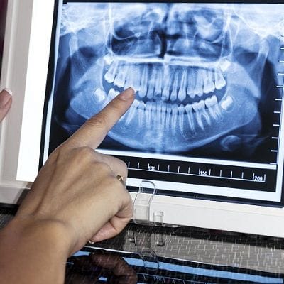
Panoramic X-Rays:
These X-beams offer a wide, far-reaching perspective on the whole oral cavity, including the teeth, jaws, sinus depressions, and temporomandibular joints (TMJ). They are useful for surveying general oral well-being, recognizing influenced teeth, finding growths or sores, and assessing the bone construction of the jaws.
Cephalometric X-Rays:
These X-beams give a side perspective of the head and neck and are normally utilized in orthodontics to survey facial and dental turn of events. Orthodontists use these rays to design orthodontic treatment, screen development, and advancement, and make exact estimations for supports and other orthodontic apparatuses.
Orthopantomogram (OPG):
Like panoramic X-beams, OPG rays give a far-reaching perspective on the upper and lower jaws, teeth, and encompassing structures. They are important for identifying affected teeth, assessing the jawbone for placement of dental implants, and surveying the general strength of the oral cavity.
Cone Bar Processed Tomography (CBCT):
CBCT is a three-layered imaging method that gives exceptionally elaborated, cross-sectional perspectives of the teeth, bones, and delicate tissues in the oral locale. It is regularly utilized in oral medical procedures, implant placement, and diagnosing complex dental circumstances.
Misconceptions:
Misinterpretations about dental X-beams are normal. It is crucial to address these errors to advance informed communication and guarantee the advantages of demonstrative imaging. Here are a few normal confusions about dental X-beams
- Dental X-rays are generally hurtful. One of the main misinterpretations is that dental X-beams are intrinsically unsafe because of radiation openness. While X-beams truly do include ionizing radiation, the sum utilized in dental X-beams is negligible and thought about as safe.
- While certain patients might find dental X-beams somewhat uncomfortable. Particularly in the event that they have a delicate gag reflex, present-day X-beam strategies, and equipment have made the cycle speedier and more agreeable.
- Some individuals may perceive that these rays cause cancer. Worries about X-beams causing malignant growth are normal. While ionizing radiation conveys some risk, the portion utilized in dental X-beams is very low and thought safe.
- Dental X-beams are not reasonable for pregnant people. While it’s prudent to limit radiation openness during pregnancy, dental X-beams are viewed as protected when important for demonstrative purposes.
Book A Consultation!
You can now effortlessly book a consultation in Dynamic Clinic in Dubai and communicate with our doctors. Filling out the consultation form is an easy approach to book your appointment. Our doctors will consult you and make you Learn the Types and Misconception About Dental X-Rays.